Imaging and Molecular Core

Shoemaker Hall houses the Imaging Core and Molecular Core, which provides students access to advanced instrumentation.
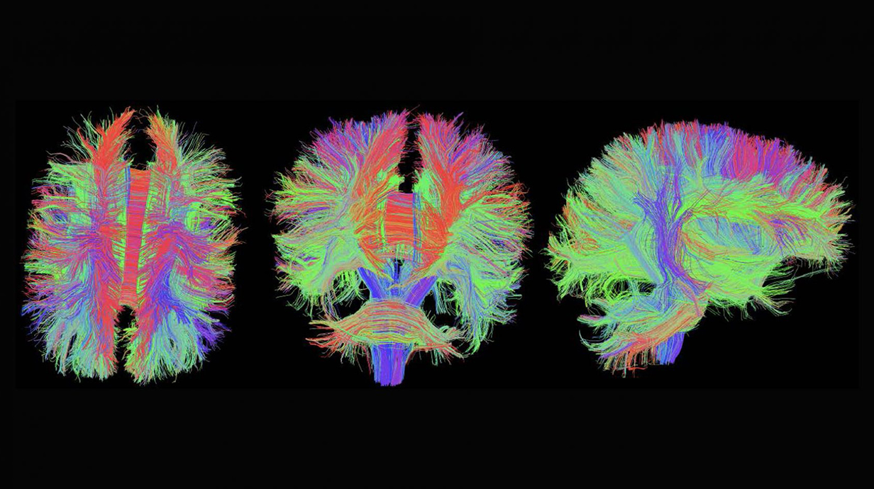
radiology brain scan, very colorful

GlyCORE Imaging Core

The objective of the GlyCORE Imaging Core is to promote and enhance the growth of glycoscience projects at the University of Mississippi, and throughout the mid-south region. The Core brings together new and existing advanced microscopes into a University-wide central platform, offering a wide range of advanced imaging techniques including laser scanning confocal microscopy and related beam parking experimental approaches, bright field and phase contrast microscopy, and computer-aided image analysis. The investigators will have access to and training in the use of advanced microscopy instrumentation, and assistance in the analysis of the resulting images. These images will provide new data and understandings on the roles of diverse carbohydrates in living systems.

As many advanced microscopic techniques are new to the University of Mississippi campus, the Imaging Core will develop a training program to inform and train new users on these new techniques. These techniques include, but are not limited to, the live cell techniques of FRET, FLIP and FRAP. Moreover, the GlyCORE Imaging Core will hold training sessions on how to properly get the most out of image analysis with instruction on the Leica Application Suite software, Image J/Fiji, the FARSIGHT toolkit for multi-dimensional microscopy, and Adobe Photoshop.

Imaging is essential for modern biological questions, and the GlyCORE Imaging Research Core will ensure glycoscience researchers at the University of Mississippi will have access to these tools. Through the availability of these instruments and training sessions, GlyCORE investigators will have a partner in advanced imaging techniques.

Prior to issuing a press release concerning any research outcome, please notify the PI of the award, Dr. Joshua Sharp, who will contact the Program Officer and NIGMS for the necessary coordination. 

Glycoscience Center of Research Excellence (GlyCORE)

Imaging Core

The Biology Imaging Core is a shared and collaborative facility with the GlyCORE Imaging Core. The facility is designed to offer a full range of biological imaging capabilities. The imaging experts provide scientific advice and technical consultation for imaging experiment design and analysis.

The core is  equipped with a Leica SP8 inverted confocal microscope system with a temperature-controlled sample chamber, a Zeiss Axio M1 upright fluorescence microscope, and a Zeiss SteREO Discovery V12 modular stereo microscope, a Flowcam cytometer, and an image analysis workstation.

The facility provides users access to advanced microscopy instrumentation, training in the microscope operation for self-service and assistance in image analysis. Contact: Dr. John Adams, Shoemaker Hall Rm 114

Imaging Core Equipment

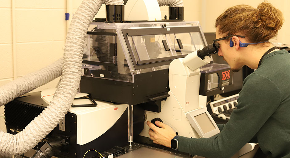
Studing using the Leica SP8 Inverted Confocal Microscope

Leica SP8 Inverted Confocal Microscope

This microscope is equipped with a motorized Super Z stage, motorize water cool turret, and 10X, 20X, 40X and 63X high NA objectives, capable of DIC and IR imaging. It is equipped with three laser systems for excitation: i) a white light laser (WWL2), ii) a 405 nm DMOD laser, and a iii) a 355 nm violet HeNe laser. This confocal also has the AOBS for tunable excitation for up to eight line simultaneously, all with very step transitions (<5nm). This is an extremely bright and spectrally flexible beam splitter that will allow for tremendous synergism with the white light laser. Also equipped with three HyD detectors and a resonant scanner, this confocal microscope is both extremely fast and sensitive. This confocal also has a powerful workstation with several software modules including: i) LAS X system for control of microscope hardware, ii) Live Data Mode, iii) 3D visualization tool, iv) 3D Analysis tools, v) Wizards for FRAP, FLIP, and FRET, and vi) Hyvolution for super resolution down to 140nm.

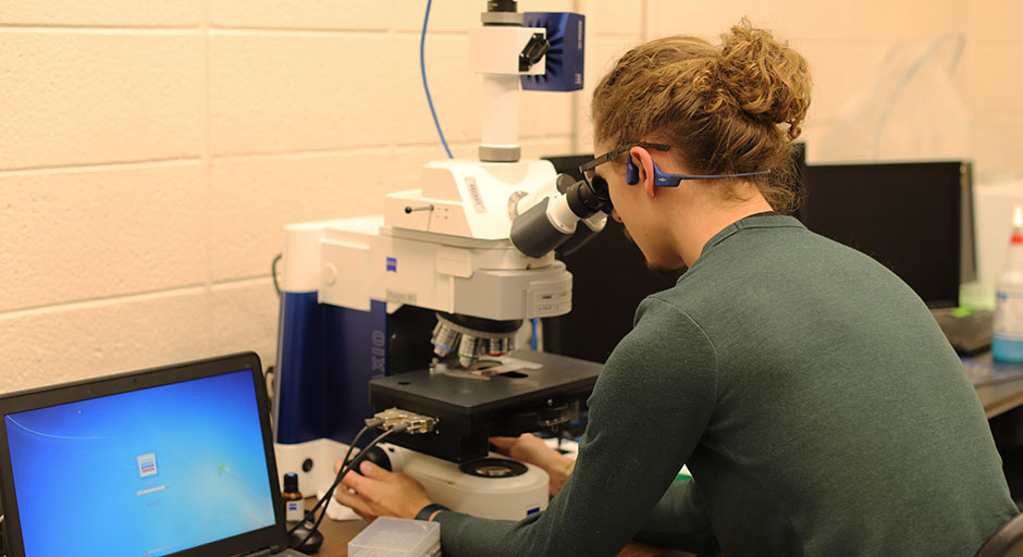
Student using the A Zeiss Axio Imager M1 microscope.

A Zeiss Axio Imager M1 Microscope

This microscope has both dark field and epifluorescent capabilities, and a mechanical stage for automated stitching. This microscope also has a 10X, 20X, 40X, and 100X Enhanced Contrast Plan NeoFluar objectives, capable of collecting high contrast images throughout the visible spectrum. The microscope has a powerful graphics workstation for image acquisition and image analysis using Axiovision software for basic morphometrics and Stereologer software (SRC Biosciences) for stereological measurements (number, length, surface, and volume).

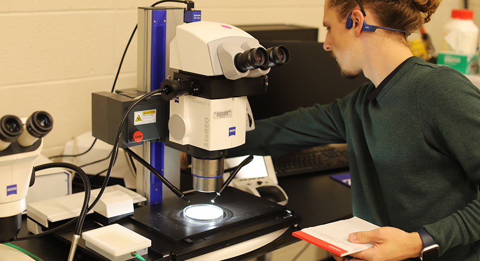
Student using A Zeiss Discovery v.12 Stereomicroscope

A Zeiss Discovery v.12 Stereomicroscope

This stereomicroscope is ideal for collecting deep macroimages at 5X to 150x magnification. It has a Plan S 1X and PlanApo S 1.5X objective, each with a large working distance and field of view. This stereomicroscope has both fluorescent and bright field light sources, and dichroic filter cubes for both GFP and RFP imaging. The stereoscope also has an Axiocam 105 color camera capable of high color reproducibility and high contrast imaging. The scope has a motorized stage, zoom, and focus controlled by its own workstation. This workstation runs ZEN 2.3 software with quantitative measurement and 3D imaging modules.

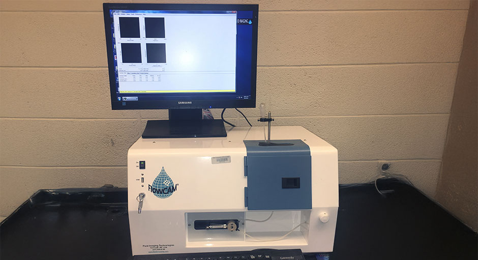
Benchtop B3 Flowcam (VS Series) equipment

Benchtop B3 Flowcam (VS Series)

The instrument is an integrated microscopic system capable of automated analysis of particles in a moving fluid. The B3 Flowcam combines capabilities of microscopy, flow cytometry, and fluorescence detection, and can automatically image, count, and analyze cells in a continuous flow.

Molecular Core

The Biology Molecular Biology Core is a self-service shared user facility that provides access to advanced instrumentation for the analysis of metabolites, proteins and nucleic acids.

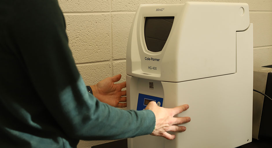
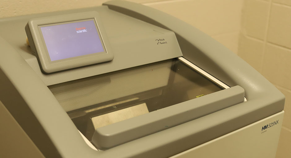
The core is equipped with an Azure C600 Advanced Imaging System for gel documentation and western blot, a Qiagen Rotor-Gene Q 5Plex real- time cycler, a QIAgility robotic system, a Biotek Synergy LX Multi-Mode Plate Reader, an OT-2 liquid handling robotic system, a Cryostat sample preparation station, and a SPEX GenoGrinder HG-400 MiniG.

Proper training is required before using any of the core instruments. Please contact the Biology Office to schedule training on any of the instruments.

Molecular Core Equipment

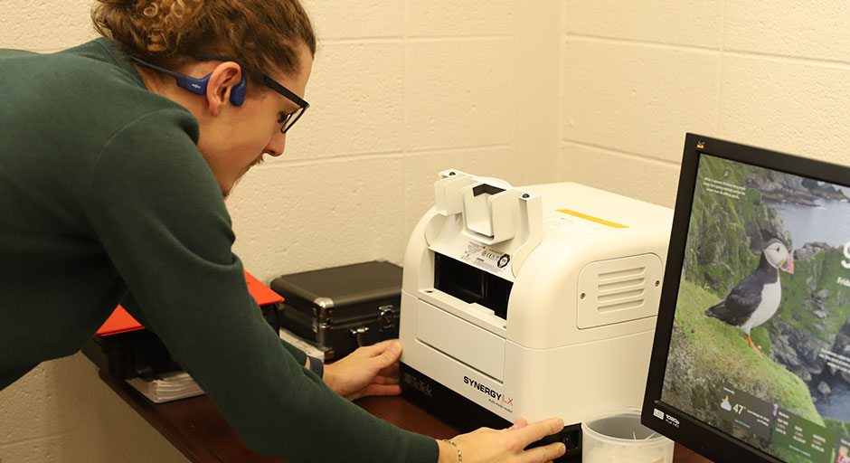
student using the Biotek Synergy LX Multi-Mode Plate Reader

Biotek Synergy LX Multi-Mode Plate Reader

This plate reader is capable of measuring UV and visible absorbance, luminescence and fluorescence intensity in a number of plate formats. The monochomator is tunable in 1 nm increments within a range from 200-999 nm. The instrument comes equipped with the Take3 plate system for low volume (2 microliters) absorption measurements, useful for quantifying RNA, DNA or Proteins. We further have red and green filter cubes for fluorescence intensity measurements. This plate reader is supported by a desktop computer running the Gen5 software.

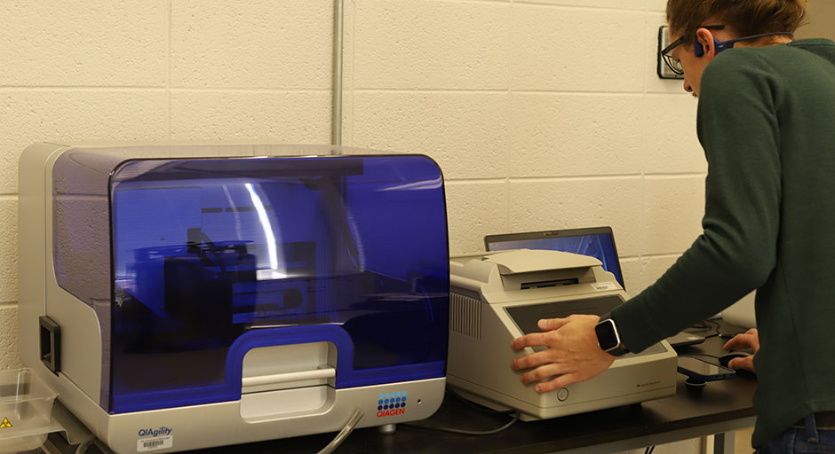
Student using the QIAgility

QIAgility

A Robot workstation for automated set up of PCR, pathogen detection, and cloning reactions. The liquid handling robot is equipped with a single channel pipetting head, and can handle a large number of tube and plate configurations with extremely high precision.

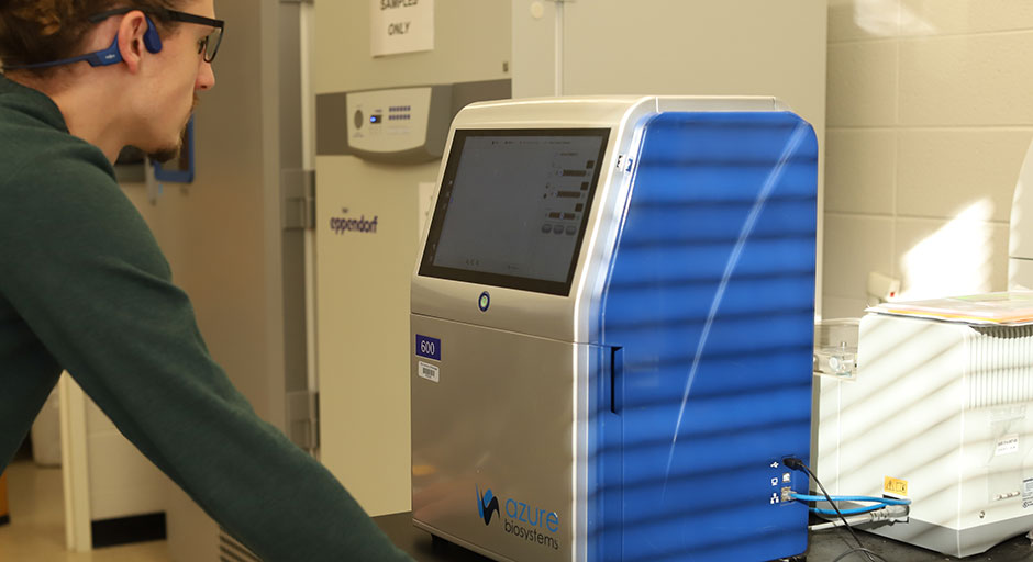
Student using the Azure C600 Advanced Imaging System

Azure C600 Advanced Imaging System

This gel documentation and Western blot system can image in the infrared fluorescence, visible fluorescence, and UV fluorescence, for DNA gels, visible fluorescent, and NIR fluorescent western blots. The imager can also capture chemiluminescence and visible dyes. The applications could even extend to colonies or seedlings on a plate. The system comes equipped with image analysis software to quantify the intensity of different bands or samples in the image.

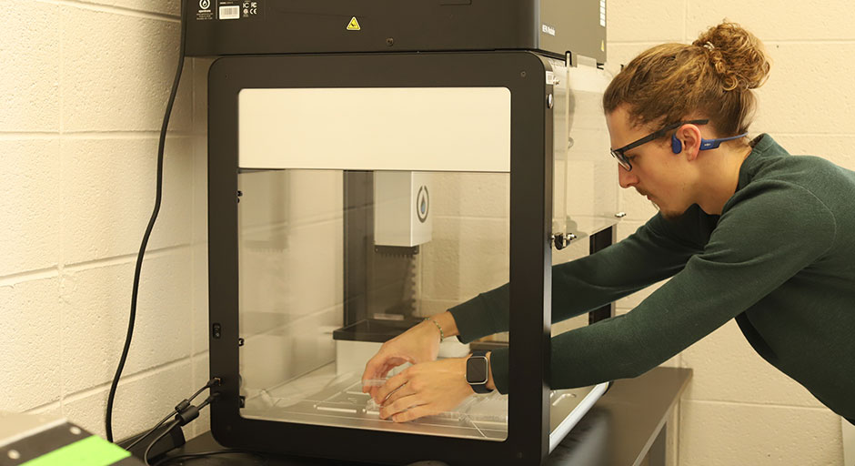
Student using the OT-2 Liquid Handling Robot

OT-2 Liquid Handling Robot

This programmable 2-armed liquid handling robot comes equipped 1 channel and an 8 channel pipette arms, with both a P50 and a P300 Multichannel pipettes. It has 11 deck slots and can fill a 96 well plate in 20 seconds. It can perform many standard tasks, reproducibly, and with minimal error. This instrument can save investigators time and money.

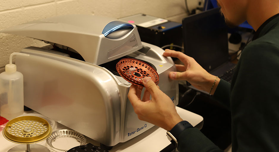
Student using the Qiagen Rotor-Gene Q 5Plex real- time cycler.

Qiagen Rotor-Gene Q 5Plex Real-Time Cycler

Student using the SPEX GenoGrinder HG-400 MiniG

SPEX GenoGrinder HG-400 MiniG

Cryostat sample preparation station

Cryostat Sample Preparation Station